Combating Mortons Neuroma

Overview

Morton neuromaMorton?s neuroma is a swollen nerve in the distal portion of the foot. The enlarged portion of the nerve represents scarring within the plantar nerve that occurs after chronic compression and/or repetitive injury. This may come about when the toes are squeezed together for too long, as can occur with the chronic use of high heels. The nerve that runs between your toes will swell and thicken. This can cause pain when walking. The symptoms of Morton?s neuroma can include burning pain in the foot, the feeling of a lump inside your foot, pain between the third and fourth toes typically but it can occur between other toes.

Causes

Although the exact cause for this condition is unclear, a number of factors can contribute to the formation of a neuroma. Biomechanical deformities, such as a high-arched foot or a flat foot, can lead to the formation of a neuroma. These foot types bring on instability around the toe joints, leading to the development of the condition. Trauma can cause damage to the nerve, resulting in inflammation or swelling of the nerve. Improper footwear that causes the toes to be squeezed together is problematic. Avoid high-heeled shoes higher than two inches. Shoes at this height can increase pressure on the forefoot area. Repeated stress, common to many occupations, can create or aggravate a neuroma.

Symptoms

Neuroma patients occasionally complain of a ?pins and needles? sensation that spreads through their feet, or of a feeling akin to hitting their ?funny bone.? The sensation may be described as similar to an electric shock. Some patients also say that these symptoms, as well as those listed above, will come and go, depending on what they are wearing on their feet, the activity they are doing, or on other external factors.

Diagnosis

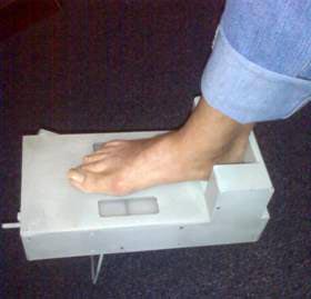
The diagnosis of a Morton’s neuroma can usually be made by the doctor when the history of pain suggests it and the examination elicits the symptoms. The foot is generally tender when the involved area is compressed and symptoms of pain and sometimes tingling can be elicited when the sides of the foot are squeezed. Magnetic resonance imaging (MRI) or ultrasound testing can be used to confirm the diagnosis if necessary.

Non Surgical Treatment

Relief of symptoms can often start by having a good pair of well fitting shoes fitted to your feet ensuring that the shoes don’t squeeze your foot together. Once footwear is addressed patients may require a small pre-metatarsal pad to be positioned onto the insole of the shoe to help lift and separate the bones in the forefoot to alleviate the pressure on the nerve. If the patients foot structure and mechanics is found to be a contributing cause, a custom made orthotic is usually the most convenient and effective way to manage the problem. Sometimes an injection of local anaesthetic and steroid is recommended to assist in settling acute symptoms.plantar neuroma

Surgical Treatment

Surgery may be considered in patients who have not responded adequately to non-surgical treatments. Your foot and ankle surgeon will determine the approach that is best for your condition. The length of the recovery period will vary, depending on the procedure performed. Regardless of whether you?ve undergone surgical or nonsurgical treatment, your surgeon will recommend long-term measures to help keep your symptoms from returning. These include appropriate footwear and modification of activities to reduce the repetitive pressure on the foot.

Prevention

It is not always possible to prevent a Morton’s neuroma. However, you probably can reduce your risk by wearing comfortable shoes that have low heels, plenty of toe space and good arch support.

The Causes Of Inferior Calcaneal Spur

Heel Spur

Overview

While the term heel spur may create the impression of a sharp bony projection on the bottom of the heel that pokes the bottom of our foot causing our pain. Painful heel spurs are actually a result of damage to the soft tissue at the bottom of the foot. While this may be confusing, we’ll try to explain. Heel spurs is the more common name for a condition that is medically referred to as plantar fascitiis or heel spur syndrome. Plantar fasciitis is a location oriented term that refers to the bottom of the foot(i.e. plantar warts). Fascia is a tough, inelastic band. ‘itis’is a term used to describe something that is inflamed (i.e. tendonitis, bursitis).

Causes

A strong band of sinew (plantar fascia) stretches across the sole of the foot below the surface of the skin and is attached to a point in the middle of the under surface of the heel bone. With repeated activity on our feet, the plantar fascia can become tight and cause persistent traction (tugging) on its attachment point into the heel bone, and inflammation and pain may develop at this site. This painful condition is known as plantar fasciitis. Sometimes a ?spur? develops at the site of this traction on the bone and protrudes into the surrounding tissue. This is a heel spur.

Posterior Calcaneal Spur

Symptoms

Some symptoms at the beginning of this condition include pain and swelling, and discomfort when pushing off with the toes during walking. This movement of the foot stretches the fascia that is already irritated and inflamed. If this condition is not treated, pain will be noticed in the heel when a heel spur develops in response to the stress. This is a common condition among athletes and others who run and jump a significant amount.

Diagnosis

A thorough medical history and physical exam by a physician is always necessary for the proper diagnosis of heel spurs and other foot conditions. X rays of the heel area are helpful, as excess bone production will be visible.

Non Surgical Treatment

There are many ways to treat heel spurs. Some remedies you can even do at home once a podiatrist shows you how. Heel spur treatment is very similar to treatment of plantar fasciitis. Here are a few of the most common treatments. First, your doctor will assess which activities are causing your symptoms and suggest rest and time off from these activities. Ice packs are used to control pain and reduce symptoms. Certain exercises and stretches help you to feel relief quickly. Medications that reduce inflammation and decrease pain are also used. Sometimes cortisone injections are given. Often special shoe orthotics can help to take the pressure off of the plantar fascia and reduce symptoms. Night splints that keep your heel stretched are sometimes recommended. Rarely, surgery is an option. A new treatment called extracorporeal shock wave therapy is being studied.

Surgical Treatment

When chronic heel pain fails to respond to conservative treatment, surgical treatment may be necessary. Heel surgery can provide relief of pain and restore mobility. The type of procedure used is based on examination and usually consists of releasing the excessive tightness of the plantar fascia, called a plantar fascia release. Depending on the presence of excess bony build up, the procedure may or may not include removal of heel spurs. Similar to other surgical interventions, there are various modifications and surgical enhancements regarding surgery of the heel.

Prevention

To prevent this condition, wearing shoes with proper arches and support is very important. Proper stretching is always a necessity, especially when there is an increase in activities or a change in running technique. It is not recommended to attempt working through the pain, as this can change a mild case of heel spurs and plantar fascitis into a long lasting and painful episode of this condition.

What Is The Perfect Solution For Inferior Calcaneal Spur

Heel Spur

Overview

Bone spurs including heel spurs occur as a natural response of the body to wear and tear. Heel spur in particular, can cause pain when it rubs against soft tissues including the Achilles tendon. When that happens movement can become restricted. Spurs can also appear in other joint areas such as under the toenail where it would lead to pain and nail deformation.

Causes

The cause of heel spurs is excessive strain placed on the plantar fascia over a long period of time, as a result of different factors. These factors include incorrect gait, being overweight, ageing or being in a job that requires a lot of standing on hard floors. It is usually a combination of any of these factors that will bring on the development of heel spurs.

Inferior Calcaneal Spur

Symptoms

Heel spurs are most noticeable in the morning when stepping out of bed. It can be described as sharp isolated pain directly below the heel. If left untreated heel spurs can grow and become problematic long-term.

Diagnosis

A Diagnosis of Heel Spur Syndrome is a very common reason for having heel pain. Heel pain may be due to other types of conditions such as tendonitis, Haglund’s Deformity, Stress Fracture, Tarsal Tunnel Syndrome, or low back problems. A more common condition in children is Sever’s Disease. The diagnosis is usually made with a combination of x-ray examination and symptoms.

Non Surgical Treatment

Ice and use arch support . If you can localize the spur, cut a hole in a pad of felt and lay the hole over the spur. This supports the area around the spur and reduces pressure on it. Massage the spur. Start gently with your thumb and gradually increase the pressure until you?re pushing hard directly on the spur with your knuckle or another firm object. Even it if hurts, it should help. Arch support. Build up an arch support system in your shoes. Try to equalize the pressure of your body weight throughout your arch and away from the plantar area. Use a ?cobra pad? or other device that supports the arch but releases pressure on the painful area. If homemade supports do not work, see a podiatrist about custom orthotics.

Surgical Treatment

In a small number of cases (usually less than 5 percent), patients may not experience relief after trying the recommendations listed above. It is important that conservative treatments (such as those listed above) be performed for AT LEAST a year before considering surgery. Time is important in curing the pain from heel spurs, and insufficient treatment before surgery may subject you to potential complications from the procedure. If these treatments fail, your doctor may consider an operation to loosen the plantar fascia, called a plantar fascia release.

Prevention

In order to prevent heel spurs, it?s important that you pay attention to the physical activities you engage in. Running or jogging on hard surfaces, such as cement or blacktop, is typical for competitive runners, but doing this for too long without breaks can lead to heel spurs and foot pain. Likewise, the shoes you wear can make a big difference in whether or not you develop heel spurs. Have your shoes and feet checked regularly by our Dallas podiatrist to ensure that you are wearing the proper equipment for the activities. Regular checkups with a foot and ankle specialist can help avoid the development of heel spurs.

What Are Bursitis Of The Foot Symptoms

Overview

A bursa is a fluid-filled sac. There are hundreds of deep and superficial bursae throughout the human body. They are typically located near major joints. Bursae are situated between bone and soft tissues like tendons, ligaments, muscles, and skin. They serve as a cushioning pad to absorb shock. The fluid within the sac is secreted to assist with friction-free movement as the soft tissues move across a bony area.

Causes

Improper foot wear, tight shoes or shoes that do not fit properly can cause extra pressure and friction on the back of the heel. Overtime, this pressure causes irritation of the bursae that protects the Achilles tendon causing one or both to swell and become inflamed. Athletes who overtrain or runners that increase their distance to quickly are at greater risk of experiencing Achilles bursitis. With over use, the Achilles bursae and tendon can become irritated and inflamed leading to thickening of the bursae lining and wearing of the tendon. Fluid builds in the bursa when it becomes irritated causing swelling of the Achilles bursa and pain at the back of the heel.

Symptoms

Pain in the heel, especially with walking, running, or when the area is touched. Pain may get worse when rising on the toes (standing on tiptoes). Red, warm skin over the back of the heel.

Diagnosis

To begin with, your doctor will gather a medical history about you and your current condition and symptoms. He/she will inquire about the level of your heel pain, the how long you have had the symptoms and the limitations you are experiencing. Details about what and when the pain started, all are very helpful in providing you with a diagnoses of your ankle / heel.

Non Surgical Treatment

Conservative treatment includes the use of shoe supports (either a heel raise or a donut-shaped heel cushion) and a limited number of local corticosteroid injections (usually up to three per year). Changing the type of footwear may be essential.

Surgical Treatment

Bursectomy is a surgical procedure used to remove an inflamed or infected bursa, which is a fluid-filled sac that reduces friction between tissues of the body. Because retrocalcaneal bursitis can cause chronic inflammation, pain and discomfort, bursectomy may be used as a treatment for the condition when it is persistent and cannot be relived with other treatments. During this procedure, a surgeon makes small incisions so that a camera may be inserted into the joint. This camera is called an arthroscope. Another small incision is made so that surgical instruments can be inserted to remove the inflamed bursa.

Prevention

To prevent bursitis of the heel in the first place, always keep proper form during exercise. In addition, don?t jump into exercises that are too intense without building up to them. Strengthen and flex your ankle.

Hammer Toe Operations

HammertoeOverview

There are two types of hammertoe. Flexible hammer toes. If the toe can still be moved at the joint, it’s a flexible hammer toe. That’s good, because this is an earlier, less-severe form of the problem. There may be several treatment options. Rigid hammer toes. If the tendons in the toe become rigid, they press the joint out of alignment. At this stage, the toe can’t be moved. This usually means surgery is required to correct the problem.

Causes

The main cause of hammer toe is poorly fitted and/or poorly designed footwear. Any footwear that is too tight in the toe box, especially high-heeled shoes, can push the toes forward, crowding one or more of them into a space that is not large enough to allow the toes to lie flat and spread as they should. Other causes include the following. Changes in foot anatomy. Sometimes the metatarsal bones in the ball of the foot can ?drop,? creating a situation in which the toes do not make contact with the surface of the shoe. The toes may then contract at one or both of the joints to re-establish contact with the surface. Traumatic injuries in which toes are jammed or broken. Diabetic neuropathy. This can cause abnormal foot biomechanics due to nerve and/or muscle damage. Damage to nerves and muscles from other conditions, such as arthritis or stroke. Heredity.

HammertoeSymptoms

A soft corn, or heloma molle, may exist in the web space between toes. This is more commonly caused by an exostosis, which is basically an extra growth of bone possibly due to your foot structure. As this outgrowth of excessive bone rubs against other toes, there is friction between the toes and a corn forms for your protection.

Diagnosis

Your doctor is very likely to be able to diagnose your hammertoe simply by examining your foot. Even before that, he or she will probably ask about your family and personal medical history and evaluate your gait as you walk and the types of shoes you wear. You’ll be asked about your symptoms, when they started and when they occur. You may also be asked to flex your toe so that your doctor can get an idea of your range of motion. He or she may hammertoe order x-rays in order to better define your deformity.

Non Surgical Treatment

In the earlier stages of hammer toe, when the toes can still be manually straightened, then conservative treatment is appropriate. This means wearing shoes which are a half size bigger than normal and which are not narrow around the toes. Exercises to stretch the toes out and strengthen the muscles under the foot which balances the tightness of the top tendons are important. Padding or corn plasters can be used to ease the discomfort of any associated corns and calluses.

Surgical Treatment

Hammer toe can be corrected by surgery if conservative measures fail. Usually, surgery is done on an outpatient basis with a local anesthetic. The actual procedure will depend on the type and extent of the deformity. After the surgery, there may be some stiffness, swelling and redness and the toe may be slightly longer or shorter than before. You will be able to walk, but should not plan any long hikes while the toe heals, and should keep your foot elevated as much as possible.

HammertoePrevention

Most cases of hammertoe can be prevented by wearing shoes that fit properly and give the toes plenty of room. Some recommended guidelines include avoid shoes with pointed or narrow toes. Avoid shoes that are too tight or short. Avoid high-heeled shoes, which can force the toes forward. If the shoes hurt, don’t wear them. Choose shoes with wide or boxy toes. Choose shoes that are a half-inch longer than your longest toe.

Can Hammertoes Cause Leg Soreness

HammertoeOverview

There are two types of hammertoes, Flexible hammertoes. If the toe still can be moved at the joint, it’s a flexible hammertoe. That’s good, because this is an earlier, milder form of the problem. There may be several treatment options. Rigid hammertoes. If the tendons in the toe become hammertoe rigid, they press the joint out of alignment. At this stage, the toe can’t be moved. It usually means that surgery is needed.

Causes

While there are a number of causes, there aren’t many specific risk factors for hammertoes, women tend to get these problems more than men, but they occur without rhyme or reason. Diabetics, however, are more likely to get a hammertoe if they have underlying nerve damage in the toes and feet.

Hammer ToeSymptoms

The symptoms of hammertoe are progressive, meaning that they get worse over time. Hammertoe causes the middle joint on the second, third, fourth, or fifth toes to bend. The affected toe may be painful or irritated, especially when you wear shoes. Areas of thickened skin (corns) may develop between, on top of, or at the end of your toes. Thickened skin (calluses) may also appear on the bottom of your toe or the ball of your foot. It may be difficult to find a pair of shoes that is comfortable to wear.

Diagnosis

First push up on the bottom of the metatarsal head associated with the affected toe and see if the toe straightens out. If it does, then an orthotic could correct the problem, usually with a metatarsal pad. If the toe does not straighten out when the metatarsal head is pushed up, then that indicates that contracture in the capsule and ligaments (capsule contracts because the joint was in the wrong position for too long) of the MTP joint has set in and surgery is required. Orthotics are generally required post-surgically.

Non Surgical Treatment

The treatment options vary with the type and severity of each hammertoe, although identifying the deformity early in its development is important to avoid surgery. Podiatric medical attention should be sought at the first indication of pain and discomfort because, if left untreated, hammertoes tend to become rigid, making a nonsurgical treatment less of an option. Your podiatric physician will examine and X-ray the affected area and recommend a treatment plan specific to your condition.

Surgical Treatment

Surgery is used when other types of treatment fail to relieve symptoms or for advanced cases of hammertoe. There are several types of surgeries to treat hammertoe. A small piece of bone may be removed from the joint (arthroplasty). The toe joint may be fused to straighten it (arthrodesis). Surgical hardware, such as a pin, may be used to hold the bones in place while they heal. Other types of surgery involve removing skin (wedging) or correcting muscles and tendons to balance the joint.

Over-Pronation Of The Foot What Are The Symptoms

Overview

Simply put, over-pronation is an excessive or poorly timed pronation which is part of the regular stride. Pronation is when the foot rolls inwards and the arch compresses as part of your normal walking motion. Pronation is necessary for shock absorption to transmit forces up and down our legs. When someone over-pronates, the foot rolls inwards to a greater degree and the arch flattens: this causes the knee and hip to come out of alignment and reduces the function of all the bones in the feet.Foot Pronation

Causes

Over-pronation occurs when the foot collapses too far inward stressing the plantar fascia (the area underneath the arch of the foot.) Normally, one pronates every time he or she walks, but excessive pronation is called over-pronation. When this occurs it can cause pain in the feet, knees, hips, low back and even the shoulder. Decreasing over-pronation, which is very prominent in runners, will help add endurance, speed and efficiency to your run and ultimately place less stress on your body.

Symptoms

Overpronation causes alterations in proper muscle recruitment patterns leading to tightness in the outside of the ankle (lateral gastrocnemius, soleus, and peroneals). This tightness can lead to weakness in the opposing muscles such as the medial gastrocnemius, anterior tibialis, and posterior tibialis. If these muscles are weak, they will not be able to keep the knee in proper alignment, causing the valgus position. All this tightness and weakness can cause pain within the ankle, calf, and knee region. And it can send imbalance and pain all the way up to the upper back, if deep core strength is lacking and can’t hold the pelvis in neutral.

Diagnosis

People who overpronate have flat feet or collapsed arches. You can tell whether you overpronate by wetting your feet and standing on a dry, flat surface. If your footprint looks complete, you probably overpronate. Another way to determine whether you have this condition is to simply look at your feet when you stand. If there is no arch on the innermost part of your sole, and it touches the floor, you likely overpronate. The only way to truly know for sure, however, is to be properly diagnosed by a foot and ankle specialist.Pronation

Non Surgical Treatment

Side Step with Opposite Reach. This exercise is designed to load the “bungee cord system” of the gluteal muscle and its opposite, latissimus dorsi muscle to keep the foot from overpronating. Because the opposite arm swings across the front leg when walking, this exercise creates tension in the muscles all the way from the front foot, across the back of the hips and back, to the fingers of the opposite hand. Movement Directions. Stand with left foot on top of the dome of the BT. (Note: For added balance, the right foot can tap on the ground, if needed). Reach right leg out to the side of the BT, and tap the ground while squatting down on the left side and reaching right arm across the left knee. Push down with left big toe while squatting. This activates the arch of the left foot and strengthens all the stabilizing muscles on the left side of the lower body. Return to starting position. Perform 8 to 10 repetitions on each leg.

Surgical Treatment

HyProCure implant. A stent is placed into a naturally occurring space between the ankle bone and the heel bone/midfoot bone. The stent realigns the surfaces of the bones, allowing normal joint function. Generally tolerated in both pediatric and adult patients, with or without adjunct soft tissue procedures. Reported removal rates, published in scientific journals vary from 1%-6%.

Diagnosing Severs Disease

Overview

If you?re a young basketballer/netballer/footballer and have heel pain when playing basketball or sports involving running or jumping, you may have a particular growth pain disorder called Sever?s Disease. It is a condition (not a disease) usually affecting 9-15 year olds that occurs at the back of the heel, where the Achilles tendon attaches to the foot. The Achilles tendon is the tendon connected to the calf muscles. Pulling of the calf muscles results in tension in the Achilles and in adolescents, repeated running/jumping can result in pain and inflammation at the heel, this is called Sever?s Disease.

Causes

There are a number of possible causes for a child’s heel pain. Because diagnosis can be challenging, a foot and ankle surgeon is best qualified to determine the underlying cause of the pain and develop an effective treatment plan. Sever’s disease, also known as calcaneal apophysitis, is by far the most common cause of heel pain in children. Other causes of heel pain include tendo-achilles bursitis, other overuse injuries, and fractures.

Symptoms

Symptoms include heel pain related to sports activities and worsen after those sport and exercise activities. However, some children who are not in a sport may also get this if they are physically active. If you notice that your child is ?walking on their toes? this is a sign of possible heel pain. The pain is usually on the back of the heel, the sides of the heel, the bottom of the heel, or a combination of all of these. We typically don’t see swelling with this, however if pressure is applied to the sides of the heel pain may be reported. Sometimes the pain is so bad the child will have to limp, or take a break from sports activity either for a few days or few months.

Diagnosis

Radiography. Most of the time radiographs are not helpful because the calcaneal apophysis is frequently fragmented and dense in normal children. But they can be used to exclude other traumas. Ultrasonography. could show the fragmentation of secondary nucleus of ossification of the calcaneus in severs?s disease. This is a safe diagnostic tool since there is no radiation. This diagnostic tool can also be used to exclude Achilles tendinitis and/or retrocalcaneal bursitis.

Non Surgical Treatment

Initially, treatment will consist of resting from activity, ice and anti-inflammatory medications to reduce the pain. Your physiotherapist may also use a variety of pain reducing techniques such as soft tissue massage or joint mobilisations. They may recommend taping to unload the area of pain, heel cups or wedge inserts into the bottom of your shoe. Also in the initial phase we may also refer you to podiatry for orthotics and/or further footwear recommendations. It is also ideal in the first instance to start stretching your calf muscles and achilles. This initial phase typically lasts for 1-2 weeks. During this time your physiotherapist will guide you on appropriate levels of activity- they may recommend you rest from impact type activities during this phase, and will guide you on the best program to return to your sport without any further injury.

What Is Adult Aquired FlatFoot ?

Overview

Adult flatfoot (adult acquired flatfoot) or posterior tibial tendon dysfunction (PTTD) is a common pathology presented to foot and ankle specialists. PTTD is characterized by a valgus (everted) hindfoot, flattening of the longitudinal arch of the foot (collapse) and abduction of the forefoot. This is a progressive deformity that begins flexible and can become rigid over time. The posterior tibial tendon (PT) is one of the main supporting structures of the foot arch. Changes within this tendon cause flattening of the foot. There are four stages of this deformity that begins flexible and progressives, with no treatment, to a rigid deformity and with time may involve the ankle joint. Patients usually present with pain in the foot or ankle stating the ankle is rolling. Its also common for patients to state they have difficulty walking barefoot. Pain is exacerbated after physical activities. Pain is usually isolated to the inside of the foot along the course of the PT tendon.Flat Feet


Causes

Obesity – Overtime if your body is carrying those extra pounds, you can potentially injure your feet. The extra weight puts pressure on the ligaments that support your feet. Also being over weight can lead to type two diabetes which also can attribute to AAFD. Diabetes – Diabetes can also play a role in Adult Acquired Flatfoot Deformity. Diabetes can cause damage to ligaments, which support your feet and other bones in your body. In addition to damaged ligaments, uncontrolled diabetes can lead to ulcers on your feet. When the arches fall in the feet, the front of the foot is wider, and outer aspects of the foot can start to rub in your shoe wear. Patients with uncontrolled diabetes may not notice or have symptoms of pain due to nerve damage. Diabetic patient don?t see they have a problem, and other complications occur in the feet such as ulcers and wounds. Hypertension – High blood pressure cause arteries narrow overtime, which could decrease blood flow to ligaments. The blood flow to the ligaments is what keeps the foot arches healthy, and supportive. Arthritis – Arthritis can form in an old injury overtime this can lead to flatfeet as well. Arthritis is painful as well which contributes to the increased pain of AAFD. Injury – Injuries are a common reason as well for AAFD. Stress from impact sports. Ligament damage from injury can cause the bones of the foot to fallout of ailment. Overtime the ligaments will tear and result in complete flattening of feet.


Symptoms

In many cases, adult flatfoot causes no pain or problems. In others, pain may be severe. Many people experience aching pain in the heel and arch and swelling along the inner side of the foot.


Diagnosis

Observe forefoot to hindfoot alignment. Do this with the patient sitting and the heel in neutral, and also with the patient standing. I like to put blocks under the forefoot with the heel in neutral to see how much forefoot correction is necessary to help hold the hindfoot position. One last note is to check all joints for stiffness. In cases of prolonged PTTD or coalition, rigid deformity is present and one must carefully check the joints of the midfoot and hindfoot for stiffness and arthritis in the surgical pre-planning.


Non surgical Treatment

Because of the progressive nature of PTTD, early treatment is advised. If treated early enough, your symptoms may resolve without the need for surgery and progression of your condition can be arrested. In contrast, untreated PTTD could leave you with an extremely flat foot, painful arthritis in the foot and ankle, and increasing limitations on walking, running, or other activities. In many cases of PTTD, treatment can begin with non-surgical approaches that may include. Orthotic devices or bracing. To give your arch the support it needs, your foot and ankle surgeon may provide you with an ankle brace or a custom orthotic device that fits into the shoe. Immobilization. Sometimes a short-leg cast or boot is worn to immobilize the foot and allow the tendon to heal, or you may need to completely avoid all weight-bearing for a while. Physical therapy. Ultrasound therapy and exercises may help rehabilitate the tendon and muscle following immobilization. Medications. Nonsteroidal anti-inflammatory drugs (NSAIDs), such as ibuprofen, help reduce the pain and inflammation. Shoe modifications. Your foot and ankle surgeon may advise changes to make with your shoes and may provide special inserts designed to improve arch support.

Adult Acquired Flat Feet


Surgical Treatment

Flatfoot reconstruction (osteotomy). This is often recommended for flexible flatfoot condition. Flatfoot reconstruction involves cutting and shifting the heel bone into a more neutral position, transferring the tendon used to flex the lesser toes (all but the big toe) to strengthen the posterior tibial tendon, and lengthening the calf muscle. Fusion (also known as triple arthrodesis). Fusion involves fusing, or making stiff, three joints in the back of the foot the subtalar, talonavicular, and calcaneocuboid joints, to realign the foot and give it a more natural shape. Pins or screws hold the area in place until it heals. Fusion is often recommended for a rigid flatfoot deformity or evidence of arthritis. Both of these surgeries can provide excellent pain relief and correction.

Do you know the Primary Treatment And Cause Of Achilles Tendon Pain ?

Overview

Achilles TendonThis nagging injury can be long-lasting if not treated – and if your running form needs some work. The name Achilles is said to be a combination of two Greek words that together mean ?grief of the people.? The injury that bears that hero?s name, in honor of his only weakness, certainly aggrieves many runners, with Achilles tendinitis accounting for around 10 percent of running injuries. Technically, Achilles tendinitis is acute inflammation of the tendon that runs along the back of the ankle. Pain in that area for longer than a couple weeks is not really tendinitis anymore. Athletes, however, tend to characterize any pain along the tendon above the back of the heel as Achilles tendinitis. Achilles tendinitis can be confused with other injuries, such as heel problems, but the hallmark sign is if you?re pinching the Achilles and it?s really sore.

Causes

Achilles tendonitis is aggravated by activities that repeatedly stress the tendon, causing inflammation. In some cases even prolonged periods of standing can cause symptoms. In many people who have developed achilles tendonitis, chronic shortening of the gastroc-soleus muscle complex is the reason that home remedies and anti-inflammatory medications fail. In these instances the muscle itself becomes shortened and creates a constant stress at the tendon?s attachment. Like a green branch that is slowly bent, eventually it begins to breakdown. Over a prolonged period the tendon becomes inflamed, and in the worst cases, appears swollen and thickened. In certain circumstances attempts to heal have failed and the body?s inability to heal the tissue results in degenerative changes known as achilles tendonosis. Anti-inflammatory medication, stretching and ice may only provide temporary relief, because they address the inflammation but not the root cause.

Symptoms

The pain associated with Achilles tendonitis can come on gradually or be caused by some type of leg or foot trauma. The pain can be a shooting, burning, or a dull ache. You can experience the pain at either the insertion point on the back of the heel or upwards on the Achilles tendon within a few inches. Swelling is also common along the area with the pain. The onset of discomfort at the insertion can cause a bump to occur called a Haglund’s deformities or Pump bump. This can be inflammation in the bursa sac that surrounds the insertion of the Achilles tendon, scar tissue from continuous tares of the tendon, or even some calcium buildup. In this situation the wearing of closed back shoes could irritate the bump. In the event of a rupture, which is rare, the foot will not be able to go through the final stage of push off causing instability. Finally, you may experience discomfort, even cramping in the calf muscle.

Diagnosis

To diagnose the condition correctly, your doctor will ask you a few questions about the pain and swelling in your heel. You may be asked to stand on the balls of your feet while your doctor observes your range of motion and flexibility. The doctor may also touch the area directly. This allows him to pinpoint where the pain and swelling is most severe.

Nonsurgical Treatment

Treatment normally includes, A bandage, designed specifically to restrict motion of the tendon. Non-steroidal anti-inflammatory medication. Orthoses (devices to help to support the muscle and relieve stress on the tendon, such as a heel pad or shoe insert. Rest, and switching to an exercise, such as swimming, that does not stress the tendon. Stretching, massage, ultrasound and appropriate exercises to strengthen the weak muscle group in front of the leg and the upper foot flexors. In extreme cases, surgery is performed, to remove fibrous tissue and repair any tears.

Achilles Tendon

Surgical Treatment

Histological and biological studies on tendon healing have made it possible to envisage surgical repair using a percutaneous approach, with the following objectives, a minimal, and not very aggressive, operation, which is quick and easy and within the capabilities of all surgeons, the shortest hospitalisation period possible, above all, early and effective re-education, providing a satisfactory result both in terms of solidity and the comfort of the patient. The percutaneous tenosynthesis TENOLIG combines stability, reliability, patient comfort and lower overall social and professional costs for this type of lesion.

Prevention

Achilles tendinitis cannot always be prevented but the following tips will help you reduce your risk. If you are new to a sport, gradually ramp up your activity level to your desired intensity and duration. If you experience pain while exercising, stop. Avoid strenuous activity that puts excessive stress on your Achilles tendon. If you have a demanding workout planned, warm up slowly and thoroughly. Always exercise in shoes that are in good condition and appropriate for your activity or sport. Be sure to stretch your calf muscles and Achilles tendon before and after working out. If you suffer from Achilles tendinitis make sure you treat it properly and promptly. If self-care techniques don?t work, don?t delay. Book a consultation with a foot care expert or you may find yourself sidelined from your favourite sports and activities.